PGIMER Orders AI-Assisted Mammography System for Breast Imaging
The Postgraduate Institute of Medical Education and Research in Chandigarh has ordered a mammography system equipped with artificial intelligence software, adding a computer-assisted layer to its breast-imaging services.

Dr Veenu Singla, professor in PGIMER’s Department of Radiodiagnosis and Imaging, confirmed the order during a press briefing on September 25, 2026. She said the technology is intended to help doctors interpret mammograms rather than make diagnoses independently.
PGIMER has not announced a delivery, installation or commissioning date. It has also not published a separate booking process, screening fee or eligibility policy for the AI-assisted service. Patients should therefore not assume that the system is already available for appointments.
What the AI is expected to do
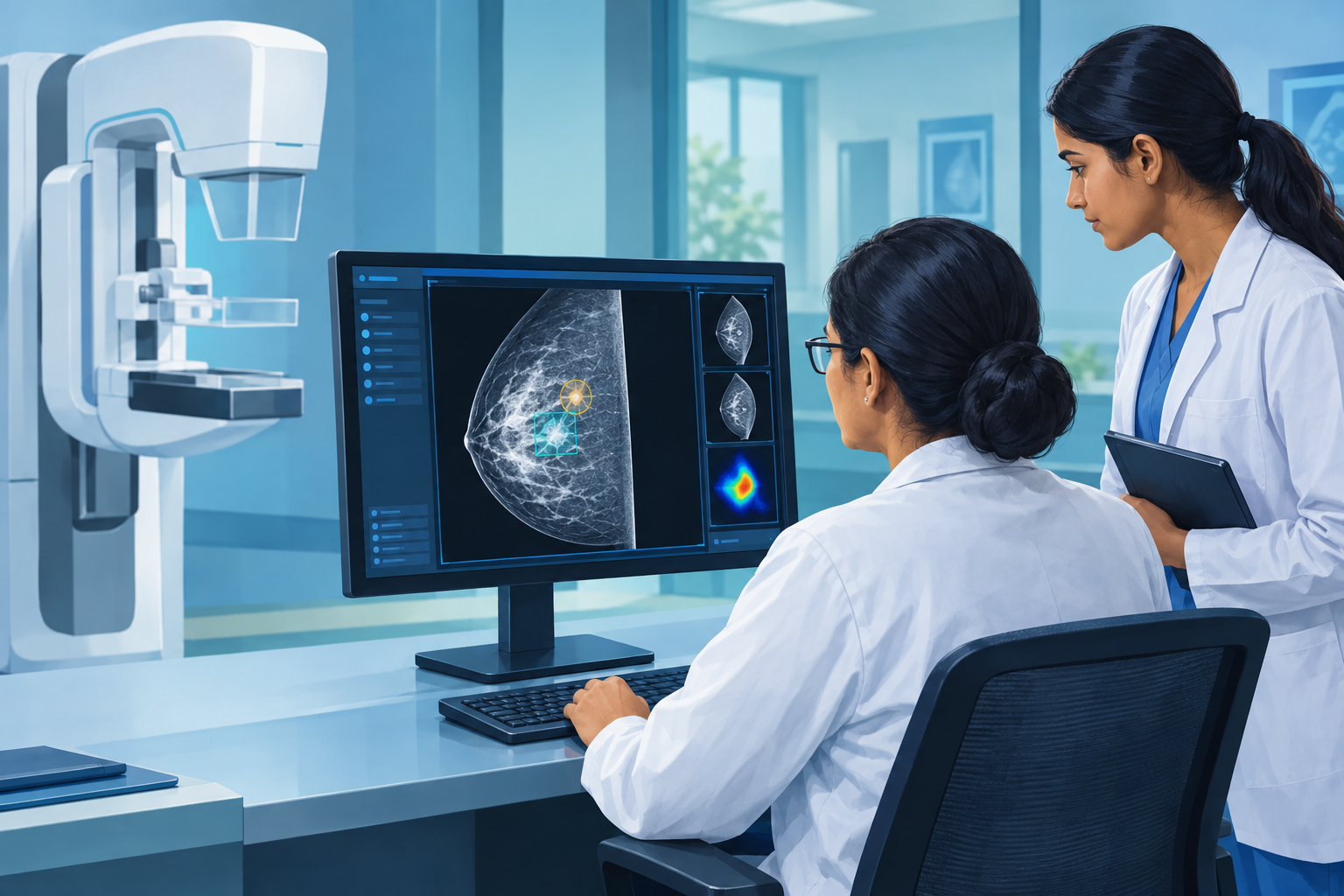
Mammography uses low-dose X-rays to produce images of breast tissue. AI software can analyse those images and draw a clinician’s attention to patterns that may require closer examination. Depending on the system, it may mark suspicious regions, estimate the likelihood of an abnormality or provide a structured assessment of breast density.
At PGIMER, one intended use is identifying small clusters of calcifications. These calcium deposits are common and are often benign, but particular patterns can be associated with early breast cancer. Subtle findings may be harder to evaluate in dense breast tissue, where normal tissue and some abnormalities can appear similar on a mammogram.
The department also expects AI to support more consistent breast-density assessment. Density is clinically relevant because it can both increase breast cancer risk and make mammograms more difficult to read. An automated measurement may give clinicians additional information, but it does not establish whether cancer is present.
Radiologists remain in charge
The software is being introduced as a decision-support tool and an additional set of checks, particularly for resident doctors learning to interpret breast images. A qualified clinician will still examine the mammogram, consider the patient’s symptoms and medical history, compare earlier images when available and decide whether any follow-up is necessary.
An AI alert is not a cancer diagnosis. A patient may need additional mammographic views, an ultrasound, follow-up imaging or a biopsy before doctors can determine what an abnormality represents. Similarly, an image not flagged by software cannot guarantee that disease is absent.
AI systems can produce false positives, which may lead to extra examinations and anxiety, as well as false negatives, in which a significant finding is not highlighted. Performance can also vary with image quality, breast density, the type of mammography equipment and how closely the patient population resembles the data used to develop the software. These limitations make clinician oversight essential.
Who may be offered mammography
PGIMER has not said that every woman attending the hospital will automatically receive AI-assisted mammography. Eligibility is expected to remain a clinical decision based on factors such as age, symptoms, examination findings, family or personal history, previous breast conditions and the purpose of the scan.
India’s public-health screening pathway recommends periodic clinical breast examination by a trained provider for women aged 30 and above. Women with a lump, nipple discharge, skin changes, persistent focal pain or another concerning finding may be referred for specialist evaluation. Mammography and ultrasound can then be selected according to age and clinical circumstances.
Women who are pregnant, may be pregnant or are breastfeeding should tell the imaging team before an examination. Younger patients and those with dense breasts may sometimes be assessed first with ultrasound, although the appropriate test depends on the individual case. People with symptoms should seek medical evaluation rather than wait for a routine screening opportunity.
Availability still to be clarified
PGIMER already provides specialised breast-imaging and intervention services, including synthesised mammography, marker placement for selected patients receiving chemotherapy and vacuum-assisted breast biopsy. The institute has said it is also exploring AI applications in ultrasound.
For the new mammography system, key operational details remain pending. PGIMER has not stated when patient scans will begin, whether the AI analysis will apply to every mammogram, or whether access will require referral through a particular outpatient unit. Those details are likely to become clearer after delivery, technical validation, staff training and integration into the hospital’s reporting workflow.
The planned upgrade may give clinicians useful additional information, but its value will depend on careful implementation, quality control and continued human review. Patients should treat the technology as an aid to the breast-imaging team, not as a replacement for radiologists, clinical examination or biopsy-based diagnosis.